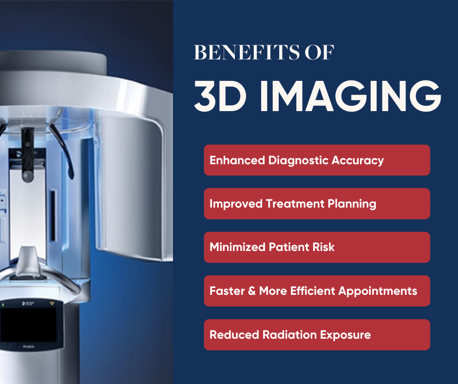
How 3D Imaging Revolutionizes Dental Implants and Diagnostics
Advancements in dental technology have transformed the way Campi Dental diagnoses and treats oral health issues. One of the most groundbreaking innovations is 3D imaging, specifically Cone Beam Computed Tomography (CBCT). This technology provides us with unparalleled precision and efficiency, particularly when planning and placing dental implants. Here’s how 3D imaging is revolutionizing dental care at Campi Dental.
Enhanced Precision in Diagnostics
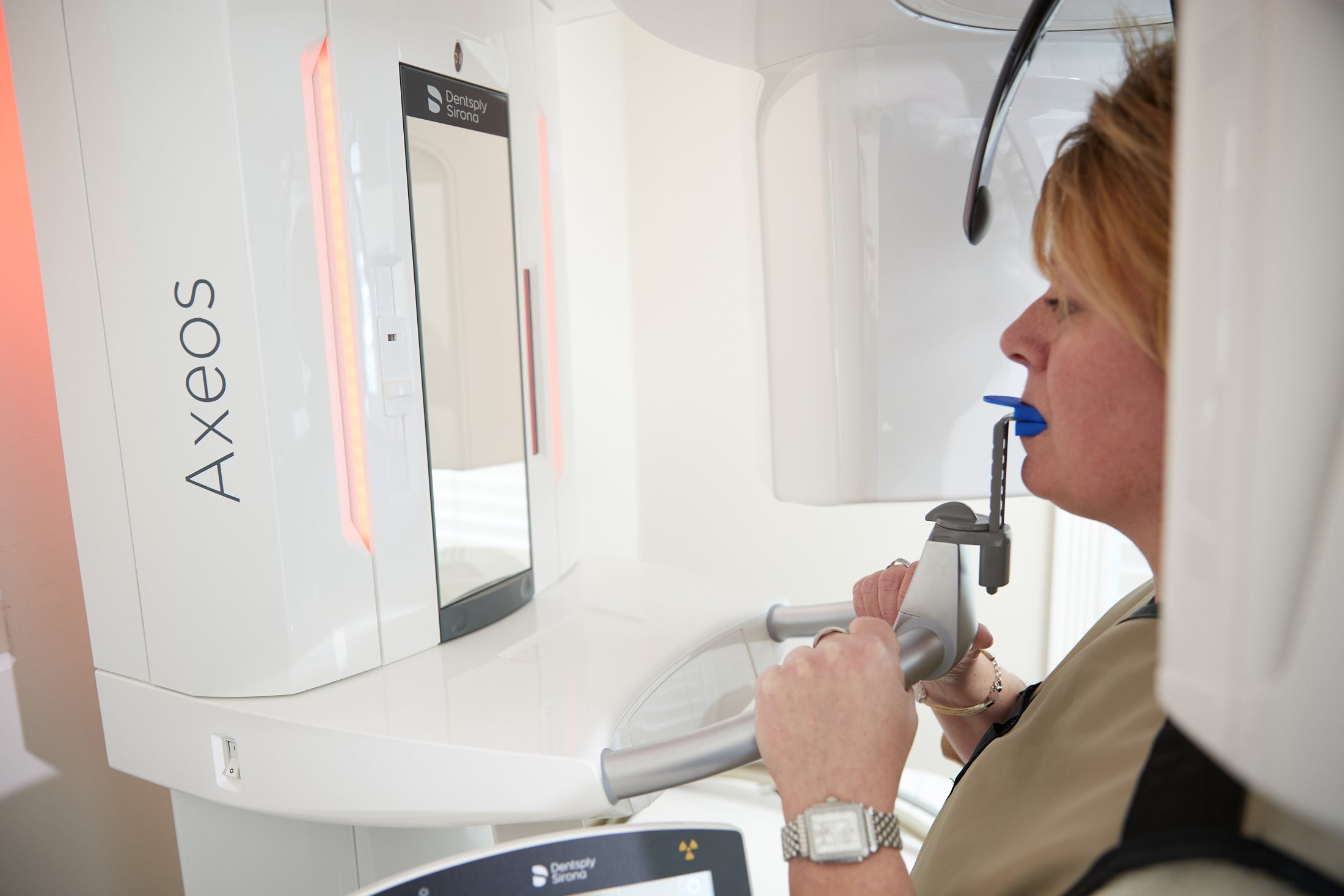
Traditional X-rays provide two-dimensional images, which can limit our ability to see the full picture of a patient’s oral structure. In contrast, 3D imaging offers highly detailed, three-dimensional views of teeth, gums, jawbone, nerves, and surrounding structures. This precision is invaluable for diagnosing complex conditions such as impacted teeth, root fractures, or bone density issues that may not be visible on traditional X-rays.
For patients considering dental implants, 3D imaging is a game-changer. It allows for an accurate assessment of the jawbone’s quality and quantity, helping determine if the patient is a candidate for implants or if additional procedures, such as bone grafting, are necessary.
Improved Implant Placement
One of the most significant benefits of 3D imaging is its role in dental implant surgery. Campi Dental can create a detailed map of the patient’s oral anatomy, including the exact location of nerves, blood vessels, and sinuses. This ensures implants are placed with pinpoint accuracy, minimizing risks and maximizing long-term success.
3D imaging allows us to deliver advanced digital treatment plans. At Campi Dental, we use the data to design surgical guides that provide a template for precise implant placement during the procedure. This reduces surgery time, enhances safety, and results in a more predictable outcome for the patient.

“3D imaging technology has truly revolutionized how we approach dental care at Campi Dental. It allows us to see specific details of a patient’s oral anatomy with unmatched precision, ensuring accurate diagnoses and improving the safety and efficiency of procedures like dental implants. By integrating this cutting-edge technology, we can provide personalized treatment plans with better outcomes, all while enhancing the overall patient experience.” – Dr. Joe Campi
Increased Patient Understanding
3D imaging isn’t just a tool for our team—it’s also beneficial for our patients. The detailed visuals make it easier for us to explain current situations, the diagnosis, and treatment plans in a way that patients can easily understand. When patients can see a 3D model of their mouth and the proposed implant placement, they’re more likely to feel confident about moving forward with treatment.
Comprehensive Applications Beyond Implants
While dental implants are a primary use for 3D imaging, its applications extend far beyond that. The Campi Dental team uses the cone beam to plan for safer and more predictable wisdom tooth extractions, assess orthodontic needs, and even detect early signs of oral cancer. Its versatility makes it an indispensable tool in modern dental care.
Why Campi Dental Invests in 3D Imaging
At Campi Dental, we are committed to providing our patients with the most advanced care possible. By incorporating 3D imaging technology, we ensure more accurate diagnoses, safer procedures, and better outcomes. Whether you’re considering dental implants or need a comprehensive evaluation, our use of cutting-edge technology ensures that you receive the best care tailored to your unique needs.
If you’re ready to experience the benefits of modern dentistry, schedule an appointment with Campi Dental today. Let us show you how 3D imaging is revolutionizing oral health care and helping us create healthier, more confident smiles!
Comments are closed.